
The University of Edinburgh
The University of Edinburgh
Delivering novel neuro-retinal biomarkers for the early diagnosis of Alzheimer’s disease
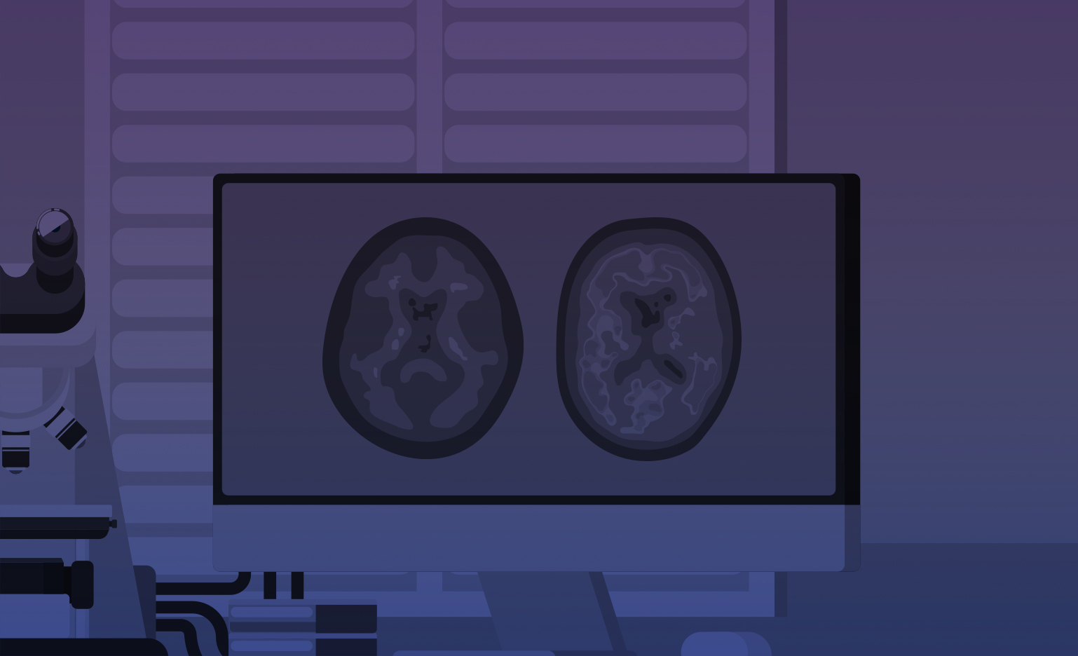
The back of the eye is the retina and it is an extension of the brain sharing a blood supply and nerve tissue. It is easily imaged by machines that are quick and comfortable for the person being examined. Most people are familiar with the technology as it is routinely used to check eye health. Our research program will look at how images from these machines can be used in new ways. Worsening health of small blood vessels in the brain is a critical feature of deteriorating brain health and declining mental ability. The health of small blood vessels in the retina is believed to closely mirror the health of similar blood vessels in the brain, which are not possible to examine with other types of routine scanning. Abnormal vessels have previously been seen in the eyes of people with Alzheimer’s disease. It is also sometimes possible to see tiny deposits or spots, called drusen, in the outlying areas of the retina. These seem to be more common in people with Alzheimer’s and are thought to reflect similar deposits forming in the brain. Another type of imaging machine shows the inner layer of the retina and its blood supply. Thinning and loss of vessels in these images suggest parallel damage to the brain in people with Alzheimer’s. Our program aims to develop the different types or modes of retinal imaging into an effective, non-invasive way of identifying early signs of Alzheimer’s through the application of computerized analysis and artificial intelligence. Ultimately, our program will enable better targeting of trials of new treatments to those who would benefit from them the most as well as helping to predict which individuals are most at risk of developing Alzheimer’s in the future, and therefore who should be closely monitored for early intervention.